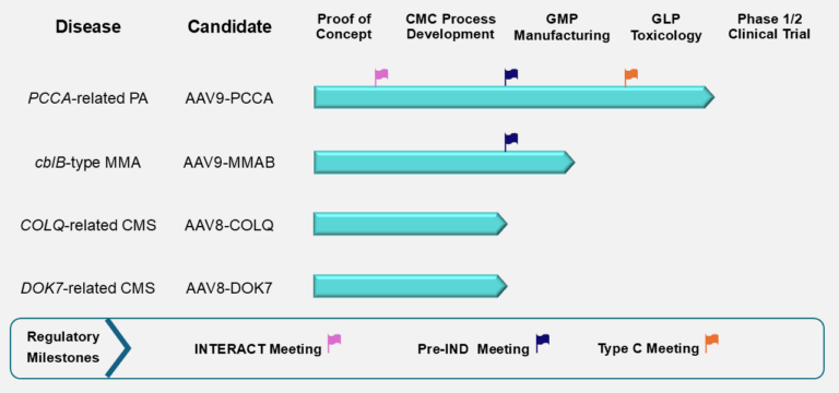
Diseases Under Study

PaVe-GT will develop gene therapies for four diseases: two congenital myasthenic syndromes and two organic acidemias. 

The congenital myasthenic syndromes under study are Dok7 deficiency and ColQ deficiency. The organic acidemias under study are propionic acidemia (caused by PCCA deficiency) and isolated methylmalonic acidemia (MMAB deficiency/cobalamin type B methylmalonic acidemia). 

Learn more about these diseases here:

Importantly, the focus of the PaVe-GT program is not on the specific diseases, but rather on increasing the efficiency of starting a gene therapy clinical trial. PaVe-GT will develop gene therapies for four diseases: two congenital myasthenic syndromes and two organic acidemias. The congenital myasthenic syndromes under study are Dok7 deficiency and ColQ deficiency. The organic acidemias under study are propionic acidemia (caused by PCCA deficiency) and isolated methylmalonic acidemia (MMAB deficiency/cobalamin type B methylmalonic acidemia). More information about these diseases is provided below.

There are many unknowns in pilot projects. They carry a high risk of failure and are likely to encounter unexpected research problems that will need to be solved. In order to increase the likelihood that the PaVe-GT approach will be successful, NIH has decided to conduct PaVe-GT using NIH investigators and facilities. This decision allows us to carry out all four trials in the same institution, and to leverage the unique resources of the NIH Clinical Center. In addition, since all personnel involved work for the federal government, we can avoid and concerns about the protection of intellectual property that could arise if the trial was carried out in a university. This is of paramount importance, given that a major goal of PaVe-GT is to put project results and documents into the public domain so that they can be made freely available to others.

These diseases were identified as suitable for PaVe-GT for the following reasons:

  • They are under study by investigators at the NIH Clinical Center.
  • They are serious diseases with no effective treatments or therapies available, such that the inherent risks of gene therapy are balanced by the potential clinical benefit. 
  • They are amenable to AAV9-mediated gene therapy. In each case, the gene is of small enough size to fit into the AAV9 vector, and cells, tissues and organs to which the gene will be delivered can be targeted by AAV9.
  • The diseases are well understood at a molecular and biochemical level.
  • Their natural histories are understood, and outcome measures have been identified.  
  • The NIH clinical investigators have years of experience with these diseases.
  • Sufficient numbers of patients are available for the clinical trials.
  • No ongoing gene therapy development for any of the disease could be identified at the beginning of the project, and given the small patient population with the diseases, future commercial interest in the disease was thought to be unlikely. 

There are no plans to add other diseases to the program. However, information developed by PaVe-GT will be shared publicly on the NIH PaVe-GT website so that everyone can benefit, and the results from PaVe-GT are intended to benefit many different gene therapy clinical trials in the future. 

The information below is technical and intended for a scientific audience. For a patient-friendly description, please visit the entry for congenital myasthenic syndromes on the Genetic and Rare Disease Information Center website. 

Congenital myasthenic syndromes (CMS) are a group of inherited disorders in which there is impairment of the highly specialized neuromuscular junction (NMJ). To date, 32 types of CMS have been described. Defects in all areas of the NMJ — including the presynaptic cell (nerve), synaptic cleft and postsynaptic cell (muscle) — have been identified as molecular causes for CMS, and most types are inherited in an autosomal recessive pattern with early onset. Although the syndromes are rare — affecting less than 9 in one million children depending on the population — the four most common recessively inherited forms of CMS result from mutations in the DOK7, COLQ, ACHE or RAPSN genes. While symptoms vary depending on the underlying cause of the disease, they may include muscle and respiratory weakness, eyelid drooping, delayed motor development, fatigue and contractures. The two forms of CMS chosen for the PaVe-GT initiative are Dok7 and ColQ deficiency. These two forms account for about 20% of CMS cases combined. Medical treatment with pyridostigmine is not usually effective. There is varied response to other treatments, such as salbutamol.

The NMJ as a specialized structure has been investigated throughout the history of neuroscience and is of paradigmatic value as a prototypical synapse. Thus, its molecular composition, morphology and physiology are well understood and researched, making it a prime candidate to study the effects of gene therapy. Additionally, these disorders of the NMJ are not primarily degenerative, in particular as the nerve and muscle integrity are concerned. Therefore, rebuilding the NMJ will restore function to the neuromuscular unit. This target is of further interest for broader applications, including ALS, sarcopenia and myasthenia gravis.

References:

Farmakidis et al. (2018). Congenital Myasthenic Syndromes: A Clinical and Treatment Approach. Current Treatment Options in Neurology. 20(9): 36.

Nicole et al. (2017). Congenital Myasthenic Syndromes or Inherited Disorders of Neuromuscular Transmission: Recent Discoveries and Open Questions. Journal of Neuromuscular Diseases. 4(4): p. 269-284.

Beeson, D. (2016). Congenital myasthenic syndromes: recent advances. Current Opinions in Neurology. 29(5): p. 565-71.

Tsai et al (2019). Congenital myasthenic syndrome in Golden Retrievers is associated with novel COLQ mutation. Journal of Veterinary Internal Medicine. 34: 258-265.

North et al (2014). Approach to the diagnosis of congenital myopathies. Neuromuscular Disorders. 24: 97-116.

Bonnemann et al (2014). Diagnostic approach to the congenital muscular dystrophies. Neuromuscular Disorders. 24: 289-311.

The information below is technical and intended for a scientific audience. For a patient-friendly description, please visit the entry for congenital myasthenic syndromes on the Genetic and Rare Disease Information Center website. 

The DOK7 gene encodes for downstream of tyrosine kinase 7 (Dok7) protein, which is essential for postsynaptic specialization of the NMJ. The primary function of the protein is to activate muscle-specific tyrosine kinase (MuSK), which subsequently results in the clustering of acetylcholine receptors in muscle cells. CMS linked to DOK7 is most often inherited recessively and may result from missense, nonsense, splice site, and/or frameshifts mutations. The most common disease causing genetic variant is the 1124_1127dupTGCC frameshift, which is located in the C-terminal domain of the protein, suggesting this region plays a significant role in its function. While many different CMS types share general phenotypic characteristics, Dok7 deficiency patients often report difficulty walking at disease onset, and some report facial, jaw and neck weakness. Fluctuation of weakness and fatigability may be less prominent compared to other forms of CMS or over longer periods of time. Thus, this condition may be mistaken for a fixed myopathy rather than a CMS, which may delay diagnosis. Although patients may experience progressive deterioration of respiratory function, eye movements are generally unaffected in these patients compared to other types of CMS. Despite this, no obvious correlation of clinical symptoms and location of mutations with the gene have been established. Current treatments available for Dok7 deficiency are pyridostigmine, ephedrine (β2-adrenergic agonist) and 3,4-diaminopyridine; however, varying responses have been reported. adeno-associated virus (AAV) gene therapy is promising for Dok7 deficiency, as a mouse model of the 1124_1127dupTGCC mutation shows restored NMJ structure and function when normal (wildtype) Dok7 is replaced.

References:

Arimura et al. (2014) DOK7 gene therapy benefits mouse models of diseases characterized by defects in the neuromuscular junction. Science. 345(6203): 1505-1508.

Hallock et al. (2010) Dok7 regulates neuromuscular synapse formation by recruiting Crk and Crk-L. Genes and Development. 24(21): 2451-2461.

Palace et al. (2007) Clinical features of the DOK7 neuromuscular junction synaptopathy. Brain. 130(6): 1507-1515.

Muller et al. (2007) Phenotypical spectrum of DOK7 mutations in congenital myasthenic syndromes. Brain. 130: 1497-1506.  

Okada et al. (2006) The muscle protein Dok7 is essential for neuromuscular synaptogenesis. Science. 312(5781): 1802-1805. 

The information below is technical and intended for a scientific audience. For a patient-friendly description, please visit the entry for congenital myasthenic syndromes on the Genetic and Rare Disease Information Center website. 

When the neurotransmitter acetylcholine is released from nerve cells, it leads to muscle contraction. Acetylcholinesterase (AChE) is concentrated at the NMJ to rapidly inactive acetylcholine after signal transmission is completed. Collagen Q, a specific nonfibrillar collagen encoded by the COLQ gene, anchors AChE in the basal lamina of the mammalian NMJ. One end of ColQ binds to AChE, and the opposite end binds to muscle-specific tyrosine kinase (MuSK) at the postsynaptic membrane. The gene has also been shown to control the development and maturation of the postsynaptic domain by regulating synaptic gene expression.

In patients with ColQ deficiency, AChE is not anchored in the synaptic cleft, therefore NMJ signaling is altered. More than 30 mutations in COLQ have been causatively linked to a CMS phenotype. Similar to Dok7 deficiency, the earliest symptoms are detected in the neonatal period or during childhood and include muscle hypotonia, fatigue, respiratory insufficiency and delayed motor development. Current treatment options include salbutamol and ephedrine, but with limited responses. A mouse model of ColQ deficiency has been produced, and the same NMJ-targeting AAV platform proposed for Dok7 deficiency could be useful for treating ColQ deficiency, as well.


References:

McMaken et al. (2019) Salbutamol modifies the neuromuscular junction in mouse model of ColQ myasthenic syndrome. Human Molecular Genetics. (28)14: 2339-2351.

Sigoillot et al. (2010) Neuromuscular junction immaturity and muscle atrophy are hallmarks of the ColQ-deficient mouse, a model of congenital myasthenic syndrome with acetylcholinesterase deficiency. FASEB. doi: 10.1096/fj.201500162.

Sigiollot et al. (2010) ColQ controls postsynaptic differentiation at the neuromuscular junction. The Journal of Neuroscience. (30)1:13-23.

The information below is technical and intended for a scientific audience. For patient-friendly descriptions of the organic acidemias under study, please visit the entries for propionic acidemia or methylmalonyl-Coenzyme A mutase deficiency on the Genetic and Rare Disease Information Center website. 

The organic acidemias (OAs) are a group of inherited metabolic disorders, which disrupt branch chain amino acid, odd-chain fatty acid and/or cholesterol metabolism and cause a buildup of organic acids in the blood (emia — medical term for blood) and urine (uria — medical term for urine). Methylmalonic acidemia (MMA), propionic acidemia (PA), isovaleric acidemia and glutaric acidemia type 1, are examples of OAs, with MMA and PA collectively representing the most common of the OAs. Typically, patients with OAs present during the newborn period with a “crisis,” most commonly in the form of a life-threatening episode of metabolic acidosis and high ammonia. The first event can be very severe and require admission to an intensive care unit, even hemodialysis. During infancy and childhood, other presentations include lethargy, vomiting, poor feeding, failure to thrive, hypotonia, seizures, developmental delay and severe illness in the setting of a mild stressor, such as a cold. Potentially lethal metabolic decompensation can occur during episodes of increased catabolism, such as illness caused by infections, stress, trauma, surgery or prolonged episodes of fasting. In the United States, most patients with OAs are detected by newborn screening. The type of OA, such as MMA or PA, can be identified by the presence of specific organic acids in blood or urine, with the exact cause determined by genetic testing. The current therapy for patients with OAs includes dietary restriction of amino acids, special formulas, carnitine supplementation and, in some patients with MMA, vitamin B12 given by injections.  Severely affected patients may receive a liver transplant in an attempt to increase the hepatic enzymatic activity. In general, the long-term clinical outcomes for OAs are poor, and there is a great need for new treatment options.

References:

Kolker S, Burgard P, Sauer SW, Okun JG. Current concepts in organic acidurias: understanding intra- and extracerebral disease manifestation. J Inherit Metab Dis. 2013;36:635–44.

Schillaci LP, DeBrosse SD, McCandless SE. Inborn Errors of Metabolism with Acidosis: Organic Acidemias and Defects of Pyruvate and Ketone Body Metabolism. Pediatr Clin North Am. 2018 Apr;65(2):209-230.

The information below is technical and intended for a scientific audience. For a patient-friendly description, please visit the entry for propionic acidemia on the Genetic and Rare Disease Information Center website. 

Propionic acidemia (PA) is a rare inherited autosomal recessive metabolic disorder caused by a deficit in propionyl-CoA carboxylase (PCC) activity. The mitochondrial localized PCC enzyme is responsible for the conversion of propionyl-CoA to D-methylmalonyl-CoA. PCC is composed of six alpha and six beta subunits encoded by the PCCA and PCCB genes. Pathogenic mutations in the PA patient population are found in the PCCA or PCCB genes at equal frequencies. Patients diagnosed with PA typically present in the early newborn period with a metabolic crisis, which can be fatal if not promptly recognized and treated. The current management of PA relies upon dietary restriction of branch chain amino acid precursors, carnitine supplementation and aggressive management during episodes of intercurrent infections and stress. Despite vigilant monitoring and proactive medical management, patients can suffer from metabolic decompensations, hyperammonemia, pancreatitis, sudden death from ventricular arrythmia, strokes of the basal ganglia, poor growth and cytopenias. The severe disease burden, high rates of morbidity and mortality and poor quality of life experienced by PA patients have led to the use of elective liver transplantation as a surgical treatment for PA to restore PCC activity and ameliorate or eliminate disease related symptoms. The use of expanded newborn screening to detect PA patients in the neonatal period adds another dimension of urgency to the development of new therapies for PA. AAV gene therapy is a potentially promising new therapy for which preclinical studies using murine models of PA caused by PCCA deficiency demonstrated a therapeutic benefit.

References:

Propionic Acidemia. Shchelochkov OA, Carrillo N, Venditti C. Editors In: Adam MP, Ardinger HH, Pagon RA, Wallace SE, Bean LJH, Stephens K, Amemiya A, editors. GeneReviews® [Internet]. Seattle (WA): University of Washington, Seattle; 1993-2020. 2012 May 17. 

Wongkittichote P, Ah Mew N, Chapman KA. Propionyl-CoA carboxylase – A review. Mol Genet Metab. 2017 Dec;122(4):145-152.

Chandler RJ, Chandrasekaran S, Carrillo-Carrasco N, Senac JS, Hofherr SE, Barry MA, Venditti CP. Adeno-associated virus serotype 8 gene transfer rescues a neonatal lethal murine model of propionic acidemia. Hum Gene Ther. 2011 Apr;22(4):477-81.

The information below is technical and intended for a scientific audience. For a patient-friendly description, please visit the entry for methylmalonyl-coenzyme A mutase deficiency on the Genetic and Rare Disease Information Center website. 

Isolated methylmalonic acidemia (MMA) is a group of heterogenous metabolic disorders caused by complete or partial deficiency of the mitochondrial enzyme methylmalonyl-CoA mutase (MMUT); a defect in the transport or synthesis of its cofactor, 5’-deoxyadenosylcobalamin; or deficiency of the enzyme methylmalonyl-CoA epimerase. These mitochondrial matrix enzymes are responsible for the metabolism of methylmalonyl-CoA to succinyl-CoA, and mutations in the genes that encode for the respective enzymes can lead to elevations of methylmalonic acid in the blood and urine. The cobalamin B type of MMA is caused by mutations in the MMAB gene and is one of the rarest forms of MMA. MMAB is one of several enzymes involved in the conversion of vitamin B12 to 5’-deoxyadenosylcobalamin, the active cofactor required for MMUT to function. The cobalamin B type of MMA is inherited in an autosomal recessive manner and can be detected by newborn screening and then confirmed by genetic testing. Patients with the cobalamin B type of MMA experience severe symptoms, which can appear in early infancy or the first year of life, of acid-base imbalance, high levels of ammonia, lethargy, vomiting, dehydration, hypotonia and delayed development. Long-term complications of MMAB MMA can include kidney disease, pancreatitis, cytopenias and neurological damage from metabolic strokes of the basal ganglia. The current treatment for MMAB MMA is dietary restriction of protein, vitamin B12 given as an injection, carnitine and intermittent antibiotics. There is no cure for the cobalamin B type of MMA. Despite medical and dietary management, patients with MMAB MMA can experience significant medical complications, underscoring the need for new therapies. Preclinical AAV gene therapy has been used successfully to treat mouse models of MMA caused by mutations in a related gene (MMUT) and, by extension, is predicted to be equally effective for the treatment of the cobalamin B type of MMA.

References:

Isolated Methylmalonic Acidemia. Manoli I, Sloan JL, Venditti CP.Editors In: Adam MP, Ardinger HH, Pagon RA, Wallace SE, Bean LJH, Stephens K, Amemiya A, editors. GeneReviews® [Internet]. Seattle (WA): University of Washington, Seattle; 1993-2020. 2005 Aug 16.

About Methylmalonic Acidemia, NHGRI Website. 

Chandler RJ, Venditti CP. Long-term rescue of a lethal murine model of methylmalonic acidemia using adeno-associated viral gene therapy. Mol Ther. 2010 Jan;18(1):11-6.

Patients and caregivers looking for information about these diseases are welcome to contact the NIH Genetic and Rare Diseases Information Center. Patients and caregivers may also find it valuable to connect with disease communities, like the Congenital Muscle Disease International Registry.

If you’d like information about enrolling in the study, please contact christopher.mendoza2@nih.gov (DOK7 deficiency or COLQ deficiency) or pastudy@mail.nih.gov (proprionic acidemia or isolated methylmalonic acidemia).


Pave-GT Resources
Rare Pediatric Disease (RPD) Designation Request for AAV9-hPCCA

This pdf file contains the RPD designation request for AAV9-hPCCA (NCATSBL-0746) and associated communications between NCATS and FDA OOPD.

Orphan Drug Designation (ODD) Request for AAV9-hPCCA

This pdf file contains the ODD request for AAV9-hPCCA (NCATSBL-0746) and associated communications between NCATS and FDA OOPD.

Subscribe
Sign-up for PaVe-GT updates.

Subscribe

Partners
National Institutes of Health